Scientific Imaging
01273 502176
Home
Contact Us


   Simply the very best in Scientific Imaging Systems

The Roslin Institute are using Camera systems and software from Digital Pixel to capture and acquire high resolution fluorescence images of various chromosome types. The system consists of a high resolution Cooled CCD camera, Pinkel filter set and and computer controlled filter wheel. This provide the theoretically best resolution on each colour channel with perfect registration and overlay characteristics.

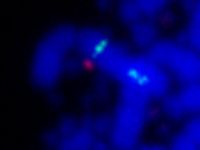
Beta-lactoglubulin transgene mRNA showing mosaic expression in mouse mammary tissue. Notice the precise registration between the two labelling channels. Image courtesy of Margaret Opsahl.


Chicken chromosomes
This image shows Fluorescence in situ hybridization of two cosmids to chicken metaphase chromosomes (tetraploid spread).
One cosmid maps to chicken chromosome 1 (green), and the other to one of the small microchromosomes (red).
Images courtesy of Jacqueline Smith.


The microchromosomes are a great example of the specific labelling that is possible with high performance camera systems, and can only be seen when the image is zoomed up, as shown here, again in green and very faintly in red.
The system has been in use for a number of years, Digital Pixel provide
a complete service contract and support solution. If you would like additional information about this system and/ or our support provision, please contact us at
support@digitalpixel.co.uk